How Is Kidney Cancer Diagnosed?

Because kidney cancer may spread to other parts of your body, it is important to be very thorough in testing for its presence. Your doctor may order some or all of a variety of tests that are available to determine the extent of your cancer and to develop your treatment plan.
Your doctor may use different approaches to diagnose the disease, depending on the symptoms you display. All approaches begin with a careful physical examination, combined with a complete discussion of past and present medical problems.
Certain tests may be done to assist your doctor in determining the correct diagnosis. The most common tests that may be ordered include:
Going to the GP:

Usually, you begin by seeing your GP who will ask you about your general health and examine you.
Your GP will ask you to give a urine sample. They will test for small amounts of blood (haematuria) which can be a sign of kidney cancer. Often the amount of blood in the urine is so small that it can't be seen but it can be picked up by the test. The doctor may also take some blood to do other tests. They may do a physical examination to feel for any lumps or swelling. But because the kidneys are deep inside the body, the doctor may not be able to feel small tumours.
Your doctor should refer you to see a specialist at the hospital if you have blood in your urine. It is important that you tell the doctor if anyone else in your family has had kidney cancer. This could help the doctor decide what tests to do.
There are guidelines for GPs to help them decide who needs an urgent referral to a specialist. These referral guidelines are covered in this section.

Lab tests:
Lab tests cannot be used to diagnose kidney cancer, but they can sometimes give the first hint that there may be a kidney problem. They are also done to get a sense of a person's overall health and to help tell if cancer may have spread to other areas. They also can help show if a person is healthy enough to have an operation.
Lab tests cannot be used to diagnose kidney cancer, but they can sometimes give the first hint that there may be a kidney problem. They are also done to get a sense of a person's overall health and to help tell if cancer may have spread to other areas. They also can help show if a person is healthy enough to have an operation.

Urinalysis:
Urinalysis (urine testing) is sometimes part of a complete physical exam, but it may not be done as a part of more routine physicals. This test may be done if your doctor suspects a kidney problem.
Microscopic and chemical tests are done on a urine sample to look for small amounts of blood and other substances not seen with the naked eye. About half of all patients with renal cell cancer will have blood in their urine. If the patient has an urothelial carcinoma (in the renal pelvis, the bladder, or other parts of the urinary tract), sometimes special microscopic examination of urine samples (called urine cytology) will show actual cancer cells in the urine.
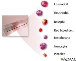
Complete blood count:
The complete blood count (CBC) is a test that measures the different cells in the blood, such as red blood cells, white blood cells, and platelets. This test result is often abnormal in people with renal cell cancer. Anemia (having too few red blood cells) is very common. Less often, a person may have too many red blood cells (called polycythemia) because the kidney cancer makes a hormone (erythropoietin) that causes the bone marrow to make more red blood cells. Blood counts are also important to make sure a person is healthy enough for surgery.
The complete blood count (CBC) is a test that measures the different cells in the blood, such as red blood cells, white blood cells, and platelets. This test result is often abnormal in people with renal cell cancer. Anemia (having too few red blood cells) is very common. Less often, a person may have too many red blood cells (called polycythemia) because the kidney cancer makes a hormone (erythropoietin) that causes the bone marrow to make more red blood cells. Blood counts are also important to make sure a person is healthy enough for surgery.

Blood chemistry tests:
Blood chemistry tests are usually done in people who might have kidney cancer, because the cancer can affect the levels of certain chemicals in the blood. For example, high levels of liver enzymes are sometimes found. High blood calcium levels may indicate that cancer has spread to the bones, and may therefore prompt a doctor to order a bone scan. Blood chemistry tests also look at kidney function, which is especially important if certain imaging tests are planned.
Blood chemistry tests are usually done in people who might have kidney cancer, because the cancer can affect the levels of certain chemicals in the blood. For example, high levels of liver enzymes are sometimes found. High blood calcium levels may indicate that cancer has spread to the bones, and may therefore prompt a doctor to order a bone scan. Blood chemistry tests also look at kidney function, which is especially important if certain imaging tests are planned.
FURTHER TESTS...!!!
If your GP refers you to a hospital specialist, further tests will help determine whether you have kidney cancer.
Going to the hospital:
The specialist will begin by asking you about your medical history and symptoms. If your urine test has picked up blood the doctor will do more urine tests. You will be asked to have more blood tests.
It is important for the doctor to take a look at your kidneys with one of the following tests
Imaging tests use x-rays, magnetic fields, or radioactive substances to create pictures of the inside of your body. Imaging tests are done for a number of reasons, including to help find out whether a suspicious area might be cancerous, to learn how far cancer may have spread, and to help determine if treatment has been effective.
Unlike most other cancers, doctors can often diagnose a kidney cancer fairly certainly without a biopsy (removal of a sample of the tumor to be looked at under a microscope). Often, imaging tests can give doctors a reasonable amount of certainty that a kidney mass is (or is not) cancerous. In some patients, however, a biopsy may be needed to be sure.
Computed tomography (CT) scans, magnetic resonance imaging (MRI) scans, and ultrasound can be very helpful in diagnosing most kinds of kidney tumors, although patients rarely need all of these tests. Other tests described here, such as chest x-rays and bone scans, are more often used to help determine if the cancer has spread (metastasized) to other parts of the body.
It is important for the doctor to take a look at your kidneys with one of the following tests
- Ultrasound
- CT scan
- Magnetic resonance imaging (MRI) scan
- Positron emission tomography (PET) scan
- Fine needle aspiration and needle core biopsy
- Angiography
- Chest x-ray
- Bone scan
Imaging tests use x-rays, magnetic fields, or radioactive substances to create pictures of the inside of your body. Imaging tests are done for a number of reasons, including to help find out whether a suspicious area might be cancerous, to learn how far cancer may have spread, and to help determine if treatment has been effective.
Unlike most other cancers, doctors can often diagnose a kidney cancer fairly certainly without a biopsy (removal of a sample of the tumor to be looked at under a microscope). Often, imaging tests can give doctors a reasonable amount of certainty that a kidney mass is (or is not) cancerous. In some patients, however, a biopsy may be needed to be sure.
Computed tomography (CT) scans, magnetic resonance imaging (MRI) scans, and ultrasound can be very helpful in diagnosing most kinds of kidney tumors, although patients rarely need all of these tests. Other tests described here, such as chest x-rays and bone scans, are more often used to help determine if the cancer has spread (metastasized) to other parts of the body.
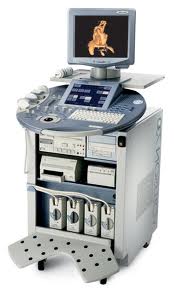
Ultrasound or ultrasonography:
If there is blood in the urine, an ultrasound of the abdomen with special attention to the kidneys, ureters, and bladder may be ordered. Usually no preparation is needed for this test.
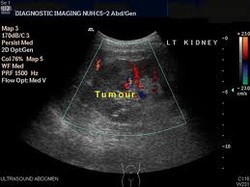
Ultrasound uses sound waves to create images of internal organs. For this test, a small, microphone-like instrument called a transducer is placed on the skin near the kidney after a gel is applied. The transducer gives off sound waves and picks up the echoes as they bounce off the tissues in the kidney. The echoes are converted by a computer into a black and white image that is displayed on a computer screen. This test is painless and does not expose you to radiation.

Ultrasound can help determine if a kidney mass is solid or filled with fluid. The echo patterns produced by most kidney tumors look different from those of normal kidney tissue. Different echo patterns also can distinguish some types of benign and malignant kidney tumors from one another. If a kidney biopsy is needed, this test can be used to guide a biopsy needle into the mass to obtain a sample.

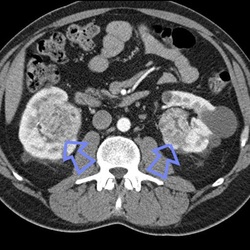
CT scan:
The computed tomography (CT or CAT) scan is an x-ray that produces detailed cross-sectional images of your body. Instead of taking one picture, like a regular x-ray, a CT scanner takes many pictures as it rotates around you while you lie on a table. A computer then combines these pictures into images of slices of the part of your body being studied.
A CT scanner has been described as a large donut, with a narrow table in the middle opening. You will need to lie still on the table while the scan is being done. CT scans will take longer than regular x-rays and you might feel a bit confined by the ring while the pictures are being taken.
Before any pictures are taken, you may be asked to drink 1 to 2 pints of a liquid called oral contrast. This helps outline the intestine so that certain areas are not mistaken for tumors. You may also receive an IV (intravenous) line through which a different kind of contrast dye (IV contrast) is injected. This helps better outline structures in your body.
The injection may cause some flushing (a feeling of warmth, especially in the face). Some people are allergic and get hives. Rarely, more serious reactions like trouble breathing or low blood pressure can occur. Be sure to tell the doctor if you have ever had a reaction to any contrast material used for x-rays.
The injection may cause some flushing (a feeling of warmth, especially in the face). Some people are allergic and get hives. Rarely, more serious reactions like trouble breathing or low blood pressure can occur. Be sure to tell the doctor if you have ever had a reaction to any contrast material used for x-rays.

CT contrast can damage the kidneys. This happens more often in patients whose kidneys are not working well in the first place. Because of this, your kidney function will be checked with a blood test before you get IV contrast.
CT scanning is one of the most useful tests for finding and looking at a tumor inside your kidney. It is also useful in checking to see if a cancer has spread to organs and tissues beyond the kidney. The CT scan will provide precise information about the size, shape, and position of a tumor, and can help find enlarged lymph nodes that might contain cancer.
CT scanning is one of the most useful tests for finding and looking at a tumor inside your kidney. It is also useful in checking to see if a cancer has spread to organs and tissues beyond the kidney. The CT scan will provide precise information about the size, shape, and position of a tumor, and can help find enlarged lymph nodes that might contain cancer.
Magnetic resonance imaging (MRI) scan:
Like CT scans, magnetic resonance imaging (MRI) scans provide detailed images of soft tissues in the body. But MRI scans use radio waves and strong magnets instead of x-rays. The energy from the radio waves is absorbed and then released in a pattern formed by the type of body tissue and by certain diseases. A computer translates the pattern into a very detailed image of parts of the body. A contrast material called gadolinium is often injected into a vein before the scan to better see details. This contrast material isn’t used in people on dialysis, because in those people it can rarely cause a severe side effect called nephrogenic systemic fibrosis.
Like CT scans, magnetic resonance imaging (MRI) scans provide detailed images of soft tissues in the body. But MRI scans use radio waves and strong magnets instead of x-rays. The energy from the radio waves is absorbed and then released in a pattern formed by the type of body tissue and by certain diseases. A computer translates the pattern into a very detailed image of parts of the body. A contrast material called gadolinium is often injected into a vein before the scan to better see details. This contrast material isn’t used in people on dialysis, because in those people it can rarely cause a severe side effect called nephrogenic systemic fibrosis.

MRI scans are a little more uncomfortable than CT scans. First, they take longer − often up to an hour. Second, you have to lie inside a narrow tube, which is confining and can upset people with claustrophobia (a fear of enclosed spaces). Special, open MRI machines can sometimes help with this if needed, but the drawback is that the pictures may not be as clear. MRI machines also make buzzing and clicking noises that many people find disturbing. Some centers provide headphones with music to block this noise out.
MRI scans are used less often than CT scans in people with kidney cancer. They may be done in cases where CT scans aren't practical, such as if a person can’t have the CT contrast dye, such as when they have an allergy to it or they don’t have good kidney function. MRI scans may also be done if there's a chance that the cancer has grown into major blood vessels in the abdomen (like the inferior vena cava), because they provide a better picture of blood vessels than CT scans. Finally, they may be used to look for possible spread of cancer to the brain or spinal cord if a person has symptoms that suggest this might be the case.
MRI scans are used less often than CT scans in people with kidney cancer. They may be done in cases where CT scans aren't practical, such as if a person can’t have the CT contrast dye, such as when they have an allergy to it or they don’t have good kidney function. MRI scans may also be done if there's a chance that the cancer has grown into major blood vessels in the abdomen (like the inferior vena cava), because they provide a better picture of blood vessels than CT scans. Finally, they may be used to look for possible spread of cancer to the brain or spinal cord if a person has symptoms that suggest this might be the case.

Positron emission tomography (PET) scan:
In a positron emission tomography (PET) scan, a form of radioactive sugar (known as fluorodeoxyglucose or FDG) is injected into the blood. The amount of radioactivity used is very low. Because cancers use glucose (sugar) at a higher rate than normal tissues, the radioactivity will tend to concentrate in the cancer. A scanner can spot the radioactive deposits and can create a picture of areas of radioactivity in the body. The picture is not finely detailed like a CT or MRI scan, but it provides helpful information about your body.
In a positron emission tomography (PET) scan, a form of radioactive sugar (known as fluorodeoxyglucose or FDG) is injected into the blood. The amount of radioactivity used is very low. Because cancers use glucose (sugar) at a higher rate than normal tissues, the radioactivity will tend to concentrate in the cancer. A scanner can spot the radioactive deposits and can create a picture of areas of radioactivity in the body. The picture is not finely detailed like a CT or MRI scan, but it provides helpful information about your body.

This test can be helpful for spotting small collections of cancer cells and can be useful in seeing if the cancer has spread to lymph nodes near the kidney. PET scans can also be useful if your doctor thinks the cancer may have spread but doesn't know where. PET scans can be used instead of several different x-rays because they scan your whole body.

Special machines can perform both a PET and CT scan at the same time (PET/CT scan). This lets the radiologist compare areas of higher radioactivity (suggesting an area of cancer) on the PET with the appearance of that area on the CT. Still, PET and PET/CT scans are not a standard part of the work-up for kidney cancers.
Fine needle aspiration and needle core biopsy:
Biopsies are not often used to diagnose kidney tumors. Imaging studies usually provide enough information for a surgeon to decide if an operation is needed. However, a biopsy is sometimes used to get a small sample of cells from an area that may be cancer when the results of imaging tests are not definite enough to warrant removing a kidney. Biopsy may also be done to confirm a cancer diagnosis if a person may not be treated with surgery, such as with small tumors that will be watched and not treated, or when other treatments are being considered.
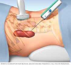
Fine needle aspiration (FNA) and needle core biopsy are 2 types of kidney biopsies that may be done. For these types of biopsies a needle is put through the skin to take a sample of cells (called per cutaneous biopsy).
For either type of biopsy, the skin where the needle is to be inserted is first numbed with local anesthesia. The doctor directs the biopsy needle into the area while looking at your kidney with either ultrasound or CT scans. Unlike ultrasound, CT doesn't provide a continuous picture, so the needle is inserted in the direction of the mass, a CT image is taken, and the direction of the needle is guided based on the image. This is repeated a few times until the needle is within the mass.
For FNA, a small sample of the target area is sucked (aspirated) through the needle into a syringe. The needle used for FNA biopsy is thinner than the ones used for routine blood tests. The needle used in core biopsies is larger than that used in FNA biopsy. It removes a small cylinder of tissue (about 1/16- to 1/8-inch in diameter and ½-inch long). Either type of sample is checked under the microscope to see if cancer cells are present.
In cases where the doctors think kidney cancer may have spread to other sites, they may take a sample of the metastatic site instead of the kidney.
Biopsies are not often used to diagnose kidney tumors. Imaging studies usually provide enough information for a surgeon to decide if an operation is needed. However, a biopsy is sometimes used to get a small sample of cells from an area that may be cancer when the results of imaging tests are not definite enough to warrant removing a kidney. Biopsy may also be done to confirm a cancer diagnosis if a person may not be treated with surgery, such as with small tumors that will be watched and not treated, or when other treatments are being considered.
Fine needle aspiration (FNA) and needle core biopsy are 2 types of kidney biopsies that may be done. For these types of biopsies a needle is put through the skin to take a sample of cells (called per cutaneous biopsy).
For either type of biopsy, the skin where the needle is to be inserted is first numbed with local anesthesia. The doctor directs the biopsy needle into the area while looking at your kidney with either ultrasound or CT scans. Unlike ultrasound, CT doesn't provide a continuous picture, so the needle is inserted in the direction of the mass, a CT image is taken, and the direction of the needle is guided based on the image. This is repeated a few times until the needle is within the mass.
For FNA, a small sample of the target area is sucked (aspirated) through the needle into a syringe. The needle used for FNA biopsy is thinner than the ones used for routine blood tests. The needle used in core biopsies is larger than that used in FNA biopsy. It removes a small cylinder of tissue (about 1/16- to 1/8-inch in diameter and ½-inch long). Either type of sample is checked under the microscope to see if cancer cells are present.
In cases where the doctors think kidney cancer may have spread to other sites, they may take a sample of the metastatic site instead of the kidney.

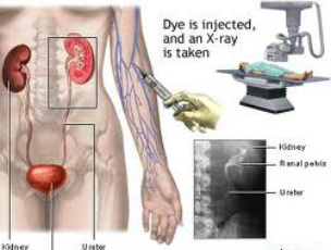
Angiography:
This procedure is used to visualize location and function of arteries.This type of x-ray also uses a contrast dye, although not the same as the one used for an IVP. A catheter is usually threaded up a large artery in your leg into the artery leading to your kidney (renal artery). The dye is then injected into the artery to identify and map the blood vessels that supply a kidney tumor. This can help in planning surgery for some patients. Angiography can also help diagnose renal cancers since the blood vessels usually have a special appearance with this test.

Angiography can be done as a part of the CT or MRI scan, instead of as a separate test. This means less contrast dye is used, which is helpful since the dye can damage kidney function further if it is given to people whose kidneys don't work as well as they should.

Chest x-ray:
If kidney cancer has been diagnosed (or is suspected), your chest may be x-rayed to see if cancer has metastasized (spread) to your lungs. Spread to the lungs is not very likely unless the cancer is far advanced. This x-ray can be done in any outpatient setting. If the results are normal, you probably don't have cancer in your lungs. The lungs are a common site of kidney cancer metastasis. Still, if your doctor has reason to suspect lung metastasis (based on symptoms like shortness of breath or a cough), you may have a chest CT scan instead of a regular chest x-ray.
If kidney cancer has been diagnosed (or is suspected), your chest may be x-rayed to see if cancer has metastasized (spread) to your lungs. Spread to the lungs is not very likely unless the cancer is far advanced. This x-ray can be done in any outpatient setting. If the results are normal, you probably don't have cancer in your lungs. The lungs are a common site of kidney cancer metastasis. Still, if your doctor has reason to suspect lung metastasis (based on symptoms like shortness of breath or a cough), you may have a chest CT scan instead of a regular chest x-ray.

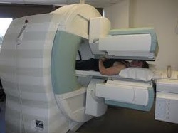
Bone scan:
A bone scan can help show whether a cancer has metastasized (spread) to your bones. For this test, a small amount of low-level radioactive material is injected into a vein (intravenously, or IV). The substance settles in areas of damaged bone throughout the entire skeleton in a couple of hours. You then lie on a table for about 30 minutes while a special camera detects the radioactivity and creates a picture of your skeleton.
A bone scan can help show whether a cancer has metastasized (spread) to your bones. For this test, a small amount of low-level radioactive material is injected into a vein (intravenously, or IV). The substance settles in areas of damaged bone throughout the entire skeleton in a couple of hours. You then lie on a table for about 30 minutes while a special camera detects the radioactivity and creates a picture of your skeleton.
Areas of active bone changes appear as "hot spots" on your skeleton − that is, they attract the radioactivity. These areas might suggest the presence of cancer spread, but arthritis or other bone diseases can also cause the same pattern. To distinguish between these conditions, your cancer care team may use other imaging tests such as simple x-rays or MRI scans to get a better look at the areas that light up, or they may even take biopsy samples of the bone.
Bone scans are done mainly when there is reason to think the cancer may have spread to the bones (like when the patient is having bone pain or blood test results show an increased calcium level). PET scans can usually show the spread of cancer to bones as well, so if you've had a PET scan you might not need a bone scan.
Bone scans are done mainly when there is reason to think the cancer may have spread to the bones (like when the patient is having bone pain or blood test results show an increased calcium level). PET scans can usually show the spread of cancer to bones as well, so if you've had a PET scan you might not need a bone scan.
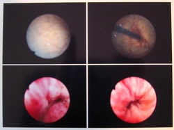
cystoscopy:
Your doctor may also want to look directly inside your bladder because this is part of the same body system as your kidneys. You might have this test if you have blood in your urine.This procedure does not inspect the kidneys, but can rule out or confirm whether any bleeding is coming from problems in the bladder.
Your doctor may also want to look directly inside your bladder because this is part of the same body system as your kidneys. You might have this test if you have blood in your urine.This procedure does not inspect the kidneys, but can rule out or confirm whether any bleeding is coming from problems in the bladder.
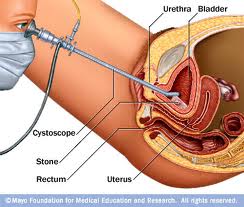
To do this test the doctor uses a cystoscope, which is a tube put into your urethra and up into the bladder. It has a light at one end and an eyepiece at the other. You can have a cystoscopy under local or general anaesthetic. You may have a cystoscopy under local anaesthetic at your first appointment because it can be done quickly and simply.
Getting your results:

Your doctor will ask you to go back to the hospital when your test results have come through. But this is bound to take a little time, even if only a few days. This can be a very anxious time.
While you are waiting for results it may help to talk to a close friend or relative about how you feel. Or you may want to contact a cancer support group to talk to someone who has been through the same experiences.
While you are waiting for results it may help to talk to a close friend or relative about how you feel. Or you may want to contact a cancer support group to talk to someone who has been through the same experiences.
